-2.jpg)
Osteoid osteoma (OO) is a small, benign bone tumor characterized by a highly vascularized core, known as the nidus, surrounded by sclerotic bone. Although it most commonly arises in the long bones, up to 20% of cases occur in the spine, where treatment can be challenging due to the proximity of nerves and other critical structures. Spinal OO typically presents with localized back or neck pain that often worsens at night.
Initial treatment is usually conservative, using aspirin or nonsteroidal anti-inflammatory drugs (NSAIDs), However, persistent symptoms often require surgical intervention, with complete removal of the nidus being essential to prevent recurrence.
Traditional surgical approaches, such as en bloc resection or curettage, can require extensive muscle dissection and bone removal, potentially compromising spinal stability and increasing the need for spinal fusion. In recent years, several minimally invasive alternatives have emerged, including radiofrequency ablation (RFA), cryoablation, microwave ablation, coagulation techniques, and MR-guided focused ultrasound. While highly effective, these approaches may be unsuitable for spinal OO when lesions are located close to neural structures because of the risk of thermal injury and the lack of histological confirmation.
A Novel Minimally Invasive Approach
A few reports have previously demonstrated the feasibility of full endoscopic resection for OO. To explore this approach further, researchers at Balgrist University Hospital investigated full endoscopic resection as a treatment for cervical and thoracic spinal OO. Published in the North American Spine Society Journal (NASSJ), the study evaluated four patients aged 22–41 years with tumors located close to neural structures, making RFA less desirable.
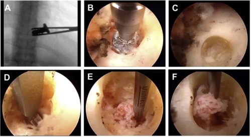
Using a minimally invasive endoscopic approach, surgeons directly visualized and completely removed the nidus while preserving surrounding tissues and spinal stability (Figure 1). Notably, this represents the first case series describing full endoscopic resection of cervical and thoracic spinal osteoid osteomas.

Excellent Clinical Outcomes
All four patients experienced immediate and complete pain relief within 24 hours after surgery, with no intraoperative or postoperative complications. Hospital stays were short (24–36 hours), blood loss was minimal, and patients were fully mobile on the first postoperative day.
Follow-up imaging demonstrated complete tumor removal with no evidence of recurrence during the follow-up period of up to 15 months (Figure 2). Histological analysis confirmed the diagnosis of osteoid osteoma in all cases (Figure 3).


Why This Matters
Full endoscopic resection offers several advantages over conventional and ablative treatments. By enabling direct visualization of the lesion, it allows complete nidus removal and histological confirmation while minimizing soft-tissue disruption and preserving spinal stability. Unlike RFA, it can be safely applied to lesions located near neural structures without the risk of thermal injury.
Although larger studies with longer follow-up are needed, the results of this first case series of cervical and thoracic spinal OO suggest that full endoscopic resection is a promising treatment option for selected patients. When performed by experienced endoscopic spine surgeons, the technique provides rapid symptom relief with minimal surgical morbidity.
Safely reaching lesions adjacent to neural structures depends on a precise spatial understanding of spinal anatomy and the surgical corridor, the foundation on which any technique, open or minimally invasive, is built.
Augmedi's interactive 3D models let residents explore exactly these anatomical relationships and surgical approaches.
At a Glance
Endoscopic Resection for Cervical and Thoracic Spinal Osteoid Osteoma
Key Collaborators
Prof. Dr. med. Mazda Farshad
Dr. med. Florian Wanivenhaus
Dr. med. José M. Spirig
Dr. med. Lukas Zingg
and colleagues
Departments and Partners
Balgrist University Hospital
Clinical Relevance
This case series demonstrated that full endoscopic resection can safely and effectively treat cervical and thoracic spinal osteoid osteomas, providing immediate pain relief, preserving spinal stability, enabling histological confirmation, and avoiding the limitations of thermal ablation techniques when lesions are located near neural structures.
Want to read the full story?
Check out the full study here:
https://www.sciencedirect.com/science/article/pii/S2666548426000284
Farshad M. et al. Endoscopic resection for cervical and thoracic spinal osteoid osteoma: A case series. North American Spine Society Journal (2026). DOI: 10.1016/j.xnsj.2026.100874.