Start every rotation with a map
When you enter a shoulder, knee, foot and ankle or another rotation, you should not have to guess what the team expects you to know. AUGMEDI organises the required knowledge into a logical path that you can follow before and during the rotation.
- A defined learning path for the rotation
- Progress from foundations to clinical practice
- A visible overview of what is complete and what remains
Learn the anatomy you will use in clinical practice
The path starts with the structures that matter for the region and then connects them to clinical anatomy. The goal is not to repeat first-year anatomy, but to recognise which structure explains the symptom, examination finding or operative risk.
- Basic and clinically applied anatomy
- Spatial relationships in realistic 3D
- Direct bridge from structure to pathology
From the patient's symptom to a structured examination
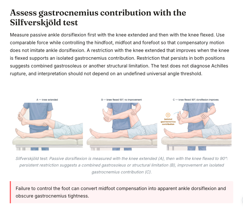
Learn the standard examination of a region, then the specific tests that answer a clinical question. Symptom-based modules connect the complaint to the relevant anatomy, examination manoeuvres and differential diagnoses so that individual facts become a clinical reasoning process.
- Standard and structure-specific examination
- Symptom-based diagnostic approach
- Interpretation of findings rather than memorising tests
Learn to read imaging in anatomical context
X-ray, CT and MRI should not be separate from the anatomy and diagnosis you have just learned. AUGMEDI connects imaging to the same structures and pathologies, while dedicated viewers can support quick orientation when you need to identify anatomy on cross-sectional imaging.
- Imaging linked to anatomy and pathology
- CT and MRI as companion tools
- Move from finding to clinical meaning
Turn findings into a treatment decision
Once the diagnosis is clear, the learning path continues to what you do next. Conservative and operative options are taught in the context of the examination, imaging and pathology that led to the decision.
- Common diagnoses and their clinical patterns
- Conservative and operative options in context
- The reasoning behind the management choice
Know the approach before you enter theatre
Surgical preparation follows the same logic: indication, positioning, landmarks, incision, deeper dissection, structures at risk and the relevant operative steps. Realistic 3D specimens help you understand the approach spatially before you see it in the operating room.
- Surgical approach broken into clear steps
- Landmarks and structures at risk made visible
- Preparation connected back to anatomy and indication
Measure your readiness before exam day
Question banks and exam simulations turn preparation into a measurable process. Instead of studying for weeks without knowing where you stand, you can test yourself, identify weak areas and direct the next study session to the topics that still need work.
- Exam-style questions with explanations
- Performance visible by topic
- Preparation focused on remaining gaps
And when you just need to look something up
Not every clinical question needs another full lesson. Atlas, Library, Cryosection, CT and MRI tools sit beside the learning path as a reference layer, so the same platform can also help when you need to check a structure or image quickly.
- Atlas and library for rapid reference
- Cryosection, CT and MRI tools
- Supporting the learning path without becoming the main story