
Not all 3D anatomy is the same.
A perfectly segmented digital muscle and a three-dimensional scan of a real human specimen can both be explored on a screen. Both can be rotated. Both are three-dimensional.
But educationally, they represent two very different views of the human body.
An idealized synthetic 3D model reduces complexity. Structures are clearly separated, consistently colored and easy to identify.
A scanned real anatomical specimen does almost the opposite. It preserves the appearance of real tissue, natural boundaries, individual variation and the complexity students will ultimately encounter in the human body.
And this distinction matters.
The relevant question for modern anatomy education may therefore no longer be:
Should we learn anatomy digitally or from real specimens?
A better question is:
How can we combine idealized 3D anatomy with photorealistic 3D anatomy to help learners move from understanding a structure to recognizing it in reality?
3D is not one learning modality
Digital anatomy is often discussed as if it were a single category. In reality, several fundamentally different representations exist.
1. Idealized synthetic 3D anatomy
Synthetic anatomical models are intentionally simplified.
Muscles can be color-coded. Nerves can be isolated. Bones can disappear with one click. Layers can be added or removed, and the learner can examine a structure without the visual interference of surrounding tissue.
This makes synthetic 3D particularly useful for building an initial mental model of anatomy.
The learner can answer fundamental questions:
- Where is the structure located?
- What lies superficial or deep to it?
- Where does it originate and insert?
- Which nerves and vessels run nearby?
- How are anatomical structures related in three-dimensional space?
Multiple studies support the educational value of interactive 3D anatomy. A systematic review and meta-analysis of randomized trials found that virtual reality anatomy teaching can improve anatomical knowledge compared with conventional approaches, although results vary substantially between studies.
2. Photorealistic 3D anatomy from real specimens
But the human body does not look like an idealized atlas.
Real muscles do not have perfectly defined red borders. Nerves may be partly hidden in connective tissue. Fascia changes the appearance of structures. Tissue color and texture vary. Anatomical relationships can be more difficult to recognize than they appear in a textbook.
This is where three-dimensional scans of real anatomical specimens provide a fundamentally different educational resource.
Techniques such as photogrammetry and structured-light scanning can transform real dissected anatomical specimens into interactive, photorealistic 3D models.
Instead of reconstructing an ideal human body, these models digitally preserve an actual anatomical specimen.
Research on photogrammetry of human specimens has shown that these models can authentically reproduce cadaveric anatomy while providing more visuospatial information than conventional two-dimensional photographs. They can also preserve anatomical details that may be simplified in computer-generated models.
This creates an important bridge between the accessibility of digital learning and the realism of specimen-based anatomy.
3. The traditional photographic atlas
Photographic atlases already address an important limitation of illustrated anatomy: they show what real anatomy looks like.
However, a photograph remains a two-dimensional representation of a three-dimensional structure.
The learner sees the specimen from the perspective selected by the photographer.
A photorealistic 3D specimen changes this interaction.
The learner can rotate the same real specimen, inspect it from different angles and explore spatial relationships that may not be evident from a single photograph.
Studies of photogrammetric anatomical models specifically highlight this ability to preserve the realism of cadaveric specimens while adding interactive three-dimensional exploration.
The educational challenge is the transition between representations
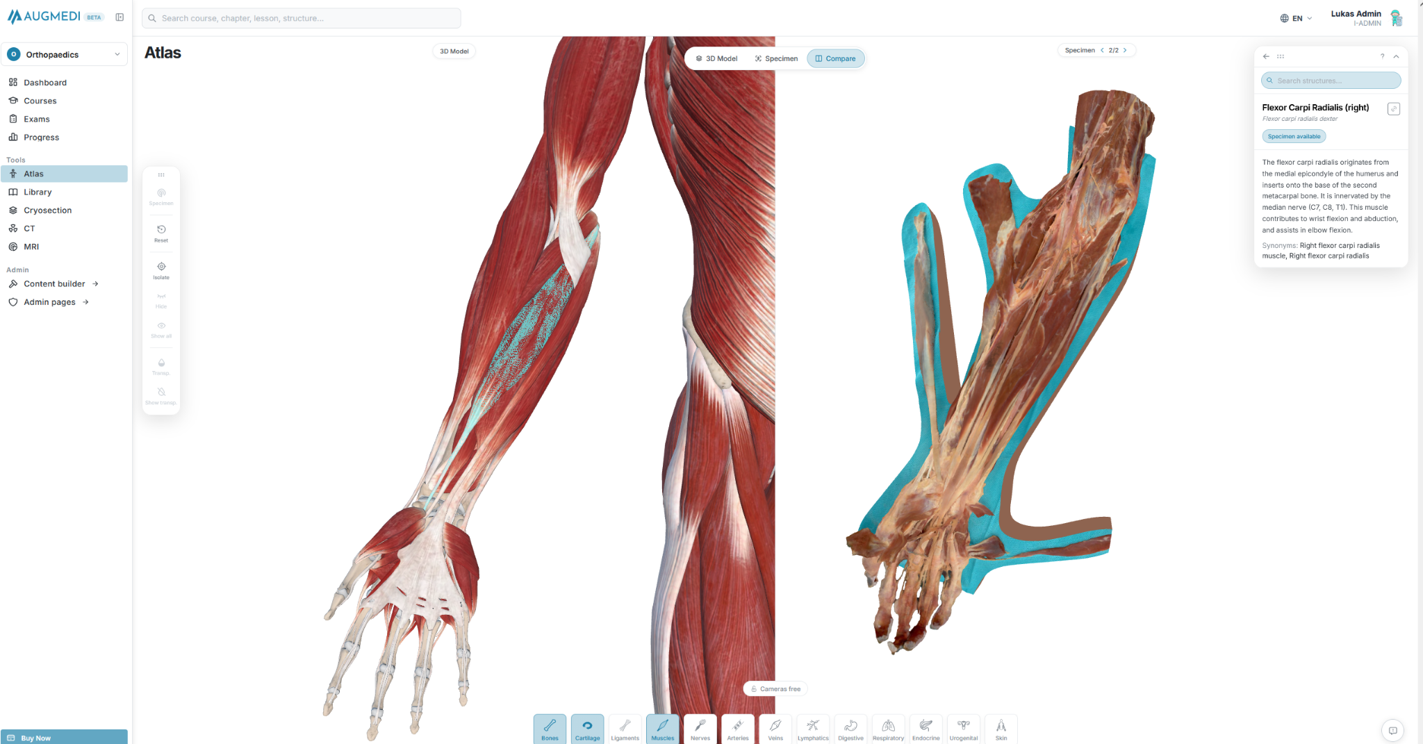
Imagine learning the flexor carpi radialis.
On an idealized model, the muscle can be highlighted in isolation. Its origin, course and tendon are immediately visible.
Now look at the same region on a real specimen.
The color is different. Fascia is present. Neighboring tendons look similar. Structures overlap. The borders are no longer perfectly defined.
The learner may understand the anatomy perfectly and still struggle to recognize it.
This is not necessarily a failure of anatomical knowledge.
It is a problem of transfer between representations.
Medical students must ultimately perform this transfer repeatedly:
Illustration → synthetic 3D model → real specimen → medical imaging → surgery → patient.
Anatomy education should therefore not only teach structures. It should train learners to recognize the same anatomy across increasingly realistic representations.
Synthetic and real 3D models solve different problems
| Representation | Main strength | Main limitation |
|---|---|---|
| Idealized synthetic 3D | Clarity, segmentation, isolation and understanding spatial relationships | Reduced biological realism and variation |
| Scanned real 3D specimen | Real tissue appearance and complexity combined with interactive 3D exploration | Structures can be more difficult to identify |
| 2D photographic atlas | Real anatomical appearance | Fixed perspective and reduced spatial exploration |
| Physical specimen | Maximum anatomical and tactile realism | Limited availability, time and repeatability |
The important conclusion is that these resources should not necessarily compete with each other.
They can form a learning sequence.
First understand. Then recognize.
For beginners, immediately confronting the full complexity of real anatomy can create unnecessary cognitive load.
An idealized model can first establish orientation.
Once the learner understands the anatomy, realism can progressively increase.
A possible progression is:
- Understand the structure in an idealized 3D model.
- Explore its anatomical relationships.
- Compare it with a scanned real human specimen.
- Recognize the structure without artificial color coding.
- Retrieve the information through active testing.
- Transfer the knowledge to imaging, surgery and clinical practice.
This is where combining different forms of digital anatomy becomes particularly interesting.
How AUGMEDI connects idealized and real 3D anatomy
This concept is built directly into the AUGMEDI Atlas.
AUGMEDI does not treat synthetic 3D anatomy and real anatomical specimens as separate worlds.
The Atlas contains both:
- structured and idealized synthetic 3D anatomy, and
- three-dimensional scans of real human anatomical specimens.
With Compare Mode, learners can place both representations directly next to each other.
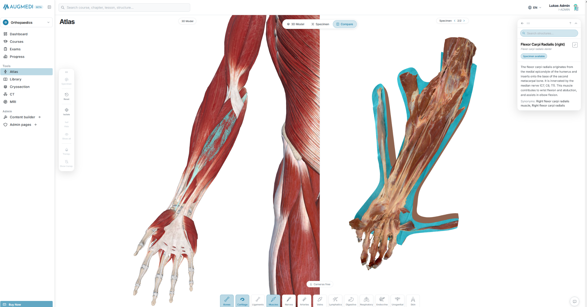
Same anatomy. Two representations.
The difference becomes immediately visible.
On one side, the learner sees the idealized anatomical representation: clean borders, distinct structures and deliberate simplification.
On the other side is a digital 3D reconstruction of an actual anatomical specimen: real tissue texture, natural anatomical boundaries and biological complexity.
Both models remain interactive and three-dimensional.
This is fundamentally different from simply placing an illustration next to a photograph.
The learner can rotate both representations, change perspective and actively investigate how the anatomy seen in the idealized model translates into the real specimen.
The objective is not just to see anatomy twice. It is to learn the relationship between abstraction and reality.
Learning should also be tested in both representations
Recognition becomes even more important during assessment.
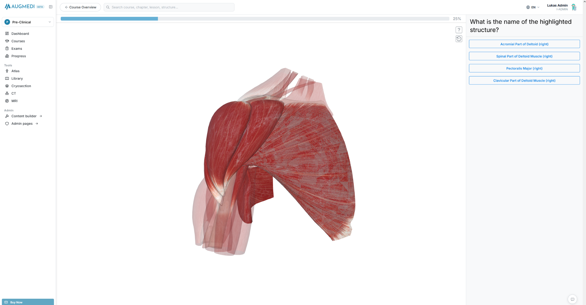
A learner who can identify a highlighted structure in an idealized model has demonstrated one level of knowledge.
Recognizing the same structure in a scanned real specimen is a different challenge.
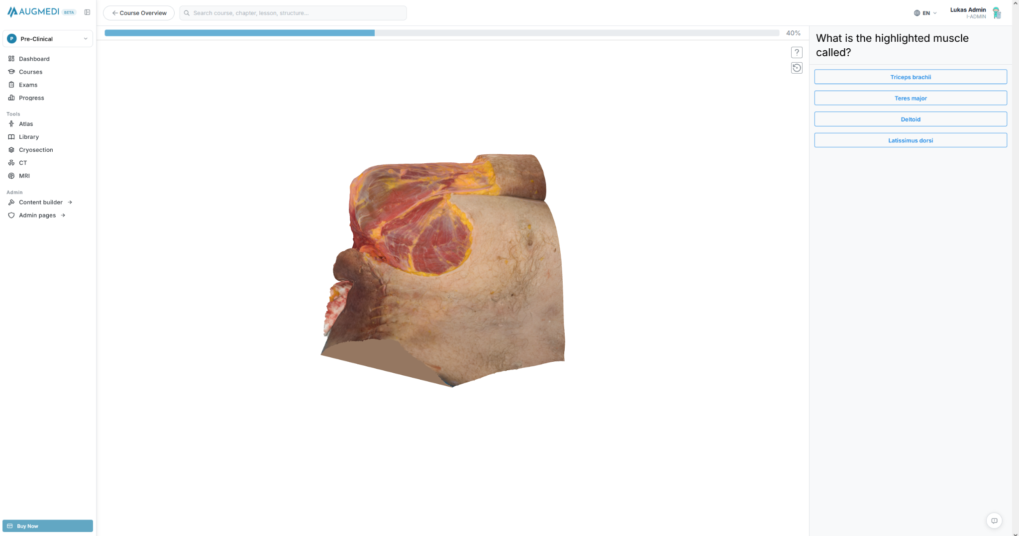
AUGMEDI can therefore use both representations not only for exploration but also for active retrieval.
This enables learners to ask a much more relevant question:
Do I only know what this structure should look like, or can I actually recognize it?
Digital anatomy should make real anatomy more valuable
The goal is not to replace cadaveric dissection or specimen-based teaching.
Physical anatomy provides experiences that no screen can completely reproduce.
But access to anatomical laboratories and specimens is inherently limited, while digital anatomy can be revisited repeatedly.
A learner can therefore prepare using an idealized model, transition to scanned real specimens and arrive at the physical anatomy laboratory with a stronger spatial framework.
Afterwards, the same digital resources can be used again for repetition and assessment.
Digital anatomy does not have to compete with real anatomy. It can prepare learners to use real anatomy more effectively.
From perfect anatomy to clinical reality
The future of anatomical education is unlikely to depend on identifying one universally superior representation.
Medicine itself requires physicians to move continuously between representations.
An anatomical structure may first be encountered in an atlas, later in a 3D model, then in a cadaver, on CT or MRI, during an operation and finally in the patient.
The educational system should prepare learners for exactly this transition.
Synthetic 3D anatomy provides clarity.
Scanned real 3D anatomy provides realism.
Medical imaging provides clinical representation.
And patients provide biological reality.
The objective should be to connect them.
At AUGMEDI, this is why we believe a modern anatomy atlas should not simply provide increasingly detailed 3D models.
It should help learners move from idealized anatomy to real anatomy and ultimately from anatomy to clinical practice.
References
- Petriceks AH, Peterson AS, Angeles M, Brown WP, Srivastava S. Photogrammetry of Human Specimens: An Innovation in Anatomy Education. Journal of Medical Education and Curricular Development. 2018;5. DOI: 10.1177/2382120518799356.
- Salimi S, Asgari Z, Mohammadnejad A, Teimazi A, Bakhtiari M. Efficacy of virtual reality and augmented reality in anatomy education: A systematic review and meta-analysis. Anatomical Sciences Education. 2024;17(9):1668–1685. DOI: 10.1002/ase.2501.
- Ail G, Freer F, Chan CS, et al. A comparison of virtual reality anatomy models to prosections in station-based anatomy teaching. Anatomical Sciences Education. 2024;17(4):763–769. DOI: 10.1002/ase.2419.
- Yun YH, Kwon HY, Jeon SK, et al. Effectiveness and satisfaction with virtual and donor dissections: A randomized controlled trial. Scientific Reports. 2024;14:16388. DOI: 10.1038/s41598-024-66292-7.
- Koucheki R, Lex JR, Morozova A, et al. Immersive virtual reality and cadaveric bone are equally effective in skeletal anatomy education: A randomized crossover noninferiority trial. Journal of Surgical Education. 2023;80:1028–1038. DOI: 10.1016/j.jsurg.2023.04.005.
- Mogali SR, Chandrasekaran R, Radzi S, et al. Investigating the effectiveness of three-dimensionally printed anatomical models compared with plastinated human specimens in learning cardiac and neck anatomy: A randomized crossover study. Anatomical Sciences Education. 2022;15(6):1007–1017. DOI: 10.1002/ase.2128.
- Radzi S, Chandrasekaran R, Peh ZK, et al. Students' learning experiences of three-dimensional printed models and plastinated specimens: a qualitative analysis. BMC Medical Education. 2022;22:695. DOI: 10.1186/s12909-022-03756-2.